Christopher (Nottingham University Hospitals) Clarke,
Anthony (University Hospitals of Leicester) Dux
John Wiley and Sons Ltd
e druk, 2015
9781118600559
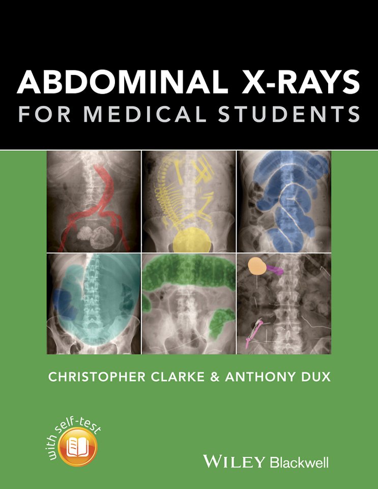
Abdominal X-rays for Medical Students
Specificaties
Paperback, 128 blz.
|
EN
John Wiley and Sons Ltd |
e druk, 2015
ISBN13: 9781118600559
Rubricering
Levertijd ongeveer 7 werkdagen
Samenvatting
Highly Commended at the British Medical Association Book Awards 2016 Abdominal X-rays for Medical Students is a comprehensive resource offering guidance on reading, presenting and interpreting abdominal radiographs.
Specificaties
ISBN13:9781118600559
Taal:EN
Bindwijze:Paperback
Aantal pagina's:128
Uitgever:John Wiley and Sons Ltd