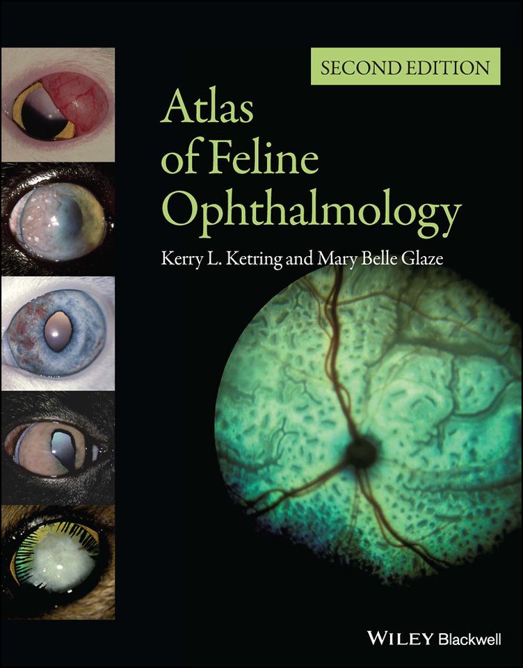
Atlas of Feline Ophthalmology
Samenvatting
Successful management of eye disease relies on the veterinarian s ability to identify ocular features and distinguish pathologic changes.
Atlas of Feline Ophthalmology, Second Edition is an invaluable diagnostic reference, providing high–quality color photographs for comparison with a presenting complaint. Presenting 394 photographs illustrating both normal and pathologic ocular conditions, this Second Edition offers a current, complete reference on ocular diseases, adding conditions recognized since publication of the first edition, a broader geographic scope, and many new images with improved quality.
Carefully designed for easy reference, the contents are divided into sections corresponding to specific anatomical structures of the eye. A useful appendix new to this edition groups figures by etiology, making it easy to find every image associated with a specific agent or disease. Atlas of Feline Ophthalmology, Second Edition is a useful tool aiding general practitioners in diagnosing eye disease in cats.
Specificaties
Inhoudsopgave
<p>Listing of Breed Predispositions to Ocular Disease page xvii</p>
<p>I. NORMAL EYE FIGURE</p>
<p>A. Diagrams</p>
<p>1. Cross–sectional 1</p>
<p>2. Fundus oculus 2</p>
<p>B. Normal adnexa/anterior segment</p>
<p>1. Frontal view 3, 4</p>
<p>2. Lateral view</p>
<p>a. Lens and cornea 5</p>
<p>b. Gross angle 6</p>
<p>3. Iridocorneal angle Gonioscopic view 7</p>
<p>C. Normal fundus 8 17</p>
<p>II. GLOBE ORBIT RELATIONSHIP</p>
<p>A. Convergent strabismus 18</p>
<p>B. Enophthalmos</p>
<p>1. Microphthalmia 19</p>
<p>2. Phthisis bulbi 20</p>
<p>3. Horner s syndrome 21</p>
<p>4. Retrobulbar tumor 22</p>
<p>5. Pain 37, 40, 42, 76, 125, 143</p>
<p>C. Exophthalmos</p>
<p>1. Cellulitis/Retrobulbar abscess 23 25</p>
<p>2. Neoplasia</p>
<p>a. Retrobulbar lymphoma 26</p>
<p>b. Zygomatic osteoma 27</p>
<p>3. Orbital pseudotumor 28, 29</p>
<p>D. Proptosis 30</p>
<p>E. Orbitalmucocele 31, 32</p>
<p>III. ADNEXA</p>
<p>A. Eyelid agenesis 33 36</p>
<p>B. Entropion 37</p>
<p>C. Ectropion 38</p>
<p>D. Distichiasis 39</p>
<p>E. Blepharitis</p>
<p>1. Herpetic 40, 54</p>
<p>2. Allergic blepharitis 41, 42, 51</p>
<p>3. Bacterial blepharitis 43</p>
<p>4. Meibomianitis 44</p>
<p>5. Demodicosis 49</p>
<p>6. Mycobacterial dermatitis 50</p>
<p>7. Food allergy 52</p>
<p>8. Pemphigus erythematosus 53</p>
<p>9. Persian idiopathic facial dermatitis 55</p>
<p>F. Apocrine cystadenoma 45, 46</p>
<p>G. Chalazion 47</p>
<p>H. Lipogranulomatous conjunctivitis 48</p>
<p>I. Granuloma/Histoplasmosis 68</p>
<p>J. Neoplasia</p>
<p>1. Cutaneous histiocytosis 56</p>
<p>2. Squamous cell carcinoma 57 59</p>
<p>3. Adenocarcinoma 60, 61</p>
<p>4. Mast cell tumor 62 64</p>
<p>5. Melanoma 65</p>
<p>6. Periorbital lymphoma 66</p>
<p>7. Nerve sheath tumor 67</p>
<p>IV. CONJUNCTIVA</p>
<p>A. Dermoid 36, 69, 70</p>
<p>B. Symblepharon 72 75, 102</p>
<p>C. Conjunctivitis</p>
<p>1. Infectious</p>
<p>a. Herpesvirus 76, 77, 83</p>
<p>b. Chlamydophila 78 80, 84</p>
<p>c. Bartonella 81, 83, 84</p>
<p>d. Mycoplasma 82</p>
<p>e. Polymicrobial 83, 84</p>
<p>f. Ophthalmia neonatorum 71</p>
<p>g. Leishmania 88</p>
<p>h. Blastomycosis 89</p>
<p>i. Histoplasmosis 90</p>
<p>2. Allergic a. Insect sting 85</p>
<p>b. Drug reaction 42</p>
<p>3. Eosinophilic 86, 87, 104, 151</p>
<p>4. Traumatic 94</p>
<p>5. Conjunctival cysts 92, 93</p>
<p>6. Parasitic–Thelaziasis 95</p>
<p>D. Dacryocystitis 96</p>
<p>E. Neoplasia</p>
<p>1. Lymphoma 91</p>
<p>2. Melanoma 97, 98</p>
<p>V. NICTITATING MEMBRANE</p>
<p>A. Nictitans protrusion</p>
<p>1. Idiopathic prolapsed nictitating membrane 99</p>
<p>2. Glandular prolapse100</p>
<p>3. Everted cartilage 101</p>
<p>4. Symblepharon 102, 113</p>
<p>5. Horner s syndrome 21</p>
<p>6. Abscess 103</p>
<p>7. Retrobulbar neoplasia 22</p>
<p>8. Phthisis bulbi 20</p>
<p>9. Pain 37, 40, 42, 76, 125, 143</p>
<p>B. Eosinophilic conjunctivitis 104</p>
<p>C. Neoplasia</p>
<p>1. Fibrosarcoma 105</p>
<p>2. Squamous cell carcinoma 106, 107</p>
<p>3. Lymphoma 108</p>
<p>4. Plasmacytoma 109</p>
<p>VI. CORNEA</p>
<p>A. Corneal opacities</p>
<p>1. Persistent pupillary membranes 110 112, 171, 172</p>
<p>2. Adherent leukoma 113, 158, 159</p>
<p>3. Corneal degeneration 114, 115</p>
<p>4. Florida spots 116, 117</p>
<p>5. Storage disease (MPS–VI)118</p>
<p>6. Relapsing polychondritis 119</p>
<p>B. Congenital Endothelial Dysfunction 123</p>
<p>C. Keratoconus 120, 121</p>
<p>D. Manx dystrophy 122</p>
<p>E. Infectious keratitis</p>
<p>1. Viral keratitis–Herpetic</p>
<p>a. Punctate 124</p>
<p>b. Dendritic 125, 126</p>
<p>c. Geographic 107, 127 131, 147, 153</p>
<p>2. Mycoplasma 132, 133</p>
<p>3. Bacterial</p>
<p>a. Staphylococcus 134</p>
<p>b. Pseudomonas135</p>
<p>4. Fungal</p>
<p>a. Candida 136</p>
<p>b. Aspergillus 137</p>
<p>5. Mycobacterial 138, 139</p>
<p>F. Ulcerative keratitis</p>
<p>1. Superficial ulceration</p>
<p>a. Keratoconjunctivitis sicca 25, 140, 153, 175, 176</p>
<p>b. Neurotrophic 140</p>
<p>2. Bullous keratitis 141</p>
<p>3. Bullous keratopathy142</p>
<p>4. Descemetocele 143, 144</p>
<p>5. Iris prolapse 35, 145</p>
<p>G. Corneal Laceration 146</p>
<p>H. Eosinophilic keratitis 107, 147 151</p>
<p>I. Corneal sequestration 37, 152 155</p>
<p>J. Foreign body 156</p>
<p>K. Staphyloma 157 159</p>
<p>L. Neoplasia</p>
<p>1. Limbal melanocytoma (Scleral shelf melanoma,</p>
<p>Epibulbar melanoma) 160 162</p>
<p>2. Neuroblastic 163</p>
<p>3. Squamous cell carcinoma 164</p>
<p>VII. ANTERIOR UVEA</p>
<p>A. Dyscorias</p>
<p>1. Iris coloboma 36, 165, 166</p>
<p>2. Corectopia 167</p>
<p>3. Idiopathic dyscoria 168</p>
<p>4. D–shaped pupil 169</p>
<p>5. Spastic pupil syndrome 170</p>
<p>B. Persistent pupillary membranes 110 112, 171, 172, 272</p>
<p>C. Chediak–Higashi syndrome 173</p>
<p>D. Iris atrophy 174</p>
<p>E. Dysautonomia 175, 176</p>
<p>F. Iris cysts/Iridocilary cysts 177 179, 267</p>
<p>G. Anterior uveitis</p>
<p>1. Iris abscess 184</p>
<p>2. Viral a. Feline leukemia complex/</p>
<p>Lymphoma 180 183, 185 190</p>
<p>b. FIV 190, 191</p>
<p>c. FIP 192 196</p>
<p>3. Toxoplasmosis 197 202</p>
<p>4. Fungal</p>
<p>a. Histoplasmosis 202 206</p>
<p>b. Cryptococcosi 207, 208</p>
<p>c. Blastomycosis 209, 210</p>
<p>d. Coccidioidomycosis 211</p>
<p>5. Bartonellosis 213 216, 244</p>
<p>6. Polymicrobial 190, 202, 212</p>
<p>7. Parasitic</p>
<p>a. Dirofilariasis 217</p>
<p>b. Myiasis 218</p>
<p>8. Metabolic/Hypertension</p>
<p>a. Hyperlipidemia 219</p>
<p>b. Systemic hypertension 220, 221</p>
<p>9. Trauma 222, 223</p>
<p>10. Lens Induced</p>
<p>a. Phacolytic 276</p>
<p>b. Septic lens implantation 224</p>
<p>11. Neoplasia</p>
<p>a. Feline diffuse iris melanoma</p>
<p>(FDIM) 225, 226, 229, 231, 269, 270</p>
<p>b. Iris melanoma 227, 228</p>
<p>c. Iris amelanotic melanoma 227, 230, 232</p>
<p>d. Iridociliary adenoma 233 236</p>
<p>e. Spindle cell tumor 237</p>
<p>f. Iridociliary leiomyoma 238</p>
<p>g. Iridociliary leiomyosarcoma 239, 240</p>
<p>h. Metastatic mammary adenocarcinoma 241</p>
<p>i. Squamous cell carcinoma242</p>
<p>j. Metastatic hemangiosarcoma 243</p>
<p>k. Primitive neural epithelial tumor244</p>
<p>l. Post–traumatic sarcoma 245, 246</p>
<p>12. Post–inflammatory sequelae</p>
<p>a. Lens capsule pigmentation 247, 250</p>
<p>b. Posterior synechia/Iris bomb´e 248, 249</p>
<p>c. Cataract 247, 248, 250</p>
<p>d. Anterior lens luxation 286</p>
<p>e. Iris cysts 179</p>
<p>f. Glaucoma 264 266, 268 270</p>
<p>VIII. GLAUCOMA</p>
<p>A. Congenital/Goniodysgenesis 251, 252, 261, 262, 288, 388</p>
<p>B. Inherited/Primary Open Angle Glaucoma (POAG)</p>
<p>1. Siamese 253</p>
<p>2. Domestic shorthair 254 257</p>
<p>C. Feline Aqueous Humor Misdirection Syndrome (FAHMS)<br /> 258 260</p>
<p>D. Secondary</p>
<p>1. Post–inflammatory/Infectious . . 209, 224, 263 265, 289</p>
<p>2. Systemic Hypertension 266</p>
<p>3. Iridocilary cysts 267</p>
<p>4. Neoplastic a. Spindle cell tumor 237</p>
<p>b. Lymphoma 190, 268</p>
<p>c. Feline diffuse iris melanoma</p>
<p>(FDIM) 232, 269, 270, 389</p>
<p>IX. LENS</p>
<p>A. Senile nuclear sclerosis 271</p>
<p>B. Cataract</p>
<p>1. Congenital/Persistent pupillary membranes 272, 274 277</p>
<p>2. Nutritional 273</p>
<p>3. Inherited 173, 275</p>
<p>4. Cataract resorption 277 279, 281</p>
<p>5. Trauma/Post–inflammatory 245 248, 280 282</p>
<p>6. Hypocalcemic 285</p>
<p>C. Cataract classification by involvement</p>
<p>1. Incipient 224, 247, 248, 258, 272, 273, 282, 285</p>
<p>2. Immature 173, 239, 247, 248, 274, 275, 280, 283</p>
<p>3. Mature 250, 260</p>
<p>4. Hypermature/Phacolytic uveitis 276, 284</p>
<p>5. Cataract resorption 19, 277 279, 281</p>
<p>D. Encephalitozoon cuniculi 283, 284</p>
<p>E. Lens luxation</p>
<p>1. Anterior 259, 260, 286, 288, 289</p>
<p>2. Posterior 254, 255, 257, 287</p>
<p>3. Subluxation 160, 256</p>
<p>X. VITREOUS</p>
<p>A. Persistent hyaloid 288</p>
<p>B. Vitreous hemorrhage 292</p>
<p>C. Hyalitis</p>
<p>1. FIV 289</p>
<p>2. Toxoplasmosis 290</p>
<p>3. Pyogranulomatous inflammation 291</p>
<p>4. Retinal detachment/Systemic hypertension 292</p>
<p>XI. RETINA AND CHOROID</p>
<p>A. Congenital</p>
<p>1. Cardiovascular Anomalies 293</p>
<p>2. Coloboma 294, 295</p>
<p>3. Retinal Folds 296, 297</p>
<p>B. Chorioretinitis–Infectious</p>
<p>1. Feline leukemia complex 298 301</p>
<p>2. Panleukopenia 302</p>
<p>3. Feline infectious peritonitis 303 308</p>
<p>4. Fungal conditions</p>
<p>a. Histoplasmosis 309 313, 386</p>
<p>b. Cryptococcosis 314 319</p>
<p>c. Blastomycosis 320 322</p>
<p>d. Coccidioidomycosis 323, 324</p>
<p>5. Toxoplasmosis 325 331</p>
<p>6. Feline hemotropic mycoplasmosis</p>
<p>(feline infectious anemia) 332</p>
<p>7. Bacterial 333</p>
<p>8. Ophthalmomyiasis 334, 335</p>
<p>C. Chorioretinitis–Traumatic 336, 349, 350</p>
<p>D. Hypertensive retinopathy 337 347</p>
<p>E. Retinal detachment</p>
<p>1. Renal failure/Systemic hypertension 338, 342 345</p>
<p>2. Trauma 348</p>
<p>3. Neoplasia 373, 374, 376</p>
<p>4. Infectious 308, 312, 313, 318, 321, 323, 331</p>
<p>F. Retinal folds</p>
<p>1. Dysplastic 296, 297</p>
<p>2. Inflammatory 305, 338</p>
<p>3. Traumatic 350</p>
<p>4. Neoplasia 371, 372</p>
<p>G. Retinopathy</p>
<p>1. Fluoroquinolone 354, 355</p>
<p>2. Feline central retinal degeneration (FCRD) 356 358</p>
<p>3. Feline generalized retinal atrophy (FGRA) 359 361</p>
<p>4. Post–trauma/Inflammation 351, 352, 387</p>
<p>5. Idiopathic 353</p>
<p>6. Progressive retinal atrophy (PRA)</p>
<p>a. Abyssinian 362, 363</p>
<p>b. Tonkinese 364</p>
<p>c. Burmese 365</p>
<p>d. Siamese 366</p>
<p>7. Chediak–Higashi syndrome 367</p>
<p>H. Vascular changes</p>
<p>1. Lipemia retinalis 368, 369</p>
<p>2. Cardiovascular anomalies293</p>
<p>3. Hyperviscosity 306</p>
<p>I. Neoplasia</p>
<p>1. Plasma cell tumor370</p>
<p>2. Retrobulbar 373, 374</p>
<p>3. Lymphoma 371, 372</p>
<p>4. Metastatic intestinal adenocarcinoma 375</p>
<p>5. Metastatic adenocarcinoma 376, 378</p>
<p>6. Metastatic hemangiosarcoma 377</p>
<p>XII. OPTIC NERVE</p>
<p>A. Coloboma 379, 380</p>
<p>B. Optic disc hypoplasia 381</p>
<p>C. Optic disc aplasia 382, 383</p>
<p>D. Optic neuritis</p>
<p>1. Cryptococcosis 319, 384</p>
<p>2. Toxoplasmosis 331</p>
<p>E. Optic nerve atrophy 385 389, 394</p>
<p>F. Glaucoma 388, 389</p>
<p>G. Neoplasia</p>
<p>1. Glioma 390</p>
<p>2. Lymphosarcoma 392</p>
<p>3. Meningioma 393, 394</p>
<p>Bibliography page 155</p>
<p>Systemic Disease Related Images page 173</p>

